
A. Protein Engineering
We use ferric hydroxamate uptake component A (FhuA), one of the members of the superfamily of bacterial outer membrane proteins that is responsible for active transport of the ferrichrome complex. Molecular design of the FhuA protein is used in biosensing, because this system exhibits a remarkable array of advantageous characteristics, including its monomeric structure, robustness, versatility, tractability, and the availability of its high-resolution crystal structure. Our strategies are aimed at developing engineered nanopore-based biosensors for bulky biopolymers, including proteins, double-stranded DNA, and their complexes with the interacting ligands.
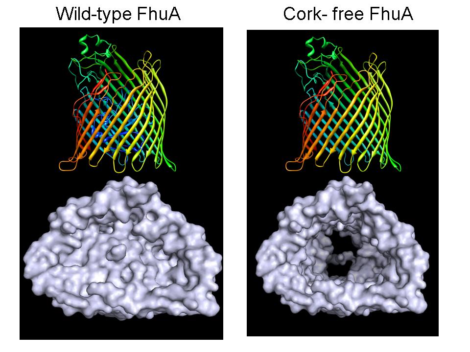
The above figure shows the wild-type (top, left-hand panel) and enginereed cork-free (top, right-handed panel) FhuA protein. The upper panels illustrate the periplasmic turns (on the bottom side of the protein) and the large cytoplasmic loops (on the top side of the protein). The lower panels represent molecular surface views of the protein from the periplasmic side.
B. Nanomedicine
The use of solid-state nanopores is at the forefront of our research in medical bionanotechnology. In analogy to their biological counterparts, solid-state nanopores are used as stochastic sensors to probe single proteins, nucleic acids and their subtle interactions with the ligands at the single-molecule level. This technique holds several advantages over its biological counterpart, including greater robustness of the pore, the ability to tune the diameter of the pore, and the potential for either integration into a lab-on-a-chip platform or parallelization for high-throughput devices. The technique also has the potential to be used as a highly sensitive and selective detection device in molecular biomedical diagnosis. Solid-State Nanopores are manufactured by our group at the Cornell Center for Materials Research (CCMR) and the Cornell Nanofabrication Facility (CNF). They are created in thin silicon nitride films by a focused electron beam. Nanopores with sizes between 2 and 20 nm are fabricated routinely.
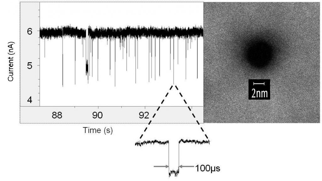
The right-hand panel illustrates a 3 nm pore in silicon nitride. Image was taken in STEM mode on the FEI Tecnai F20 TEM/STEM at the Cornell Center for Materials Research (CCMR). Special thanks go to John Grazul for his help in making an imaging of the pores. The left-hand panel shows a single-channel electrical recording of transient current blockades produced by individual double-stranded DNA.
The above single-nanopore current trace was collected by David Niedzwiecki.